Research
Mass spectrometry-based chemoproteomics is a rapidly growing field that develops and applies new technologies in areas in many disciplines including chemical biology, analytical chemistry, biochemistry, and drug development. We seek to develop and apply state of the art chemoproteomic methods to address interesting mechanistic and mode of action questions. Areas that the lab has already made substantial contributions in are summarized here.
Projects:
|
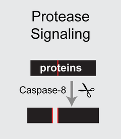
I. Towards closing the druggability gap by improving cysteine chemoproteomics and developing new electrophiles. A first key contribution of our group has been to build chemoproteomic and computational approaches that more effectively sample the entire cysteinome. We developed the SP3-FAIMS chemoproteomics platform to improve cysteine chemoproteomic coverage (ChemBioChem. 2021). To help facilitate adoption of this methodology by the community, we also authored a protocol paper describing the SP3 chemoproteomic method in detail (Desai, 2023, Current Protocols in Chemical Biology). Enabled by these enhanced screening methods, we have identified and characterized a number of novel electrophile classes—A recent example is our report of electrophiles that inactivate caspase-2 via targeting a conserved non-catalytic cysteine residue (JACS 2024).
|
|
|
Multi-omic approaches to pinpoint functional and therapeutically relevant genetic variants. Genetic variation gives rise to diversity and disease. We have established a novel chemoproteogenomics method (BioRxiv 2023) and its application to capture and perform small molecule screening at acquired cysteine single amino acid variants (SAAVs) in cancer (Fig. 1C). This study includes a GUI-enabled two-stage false discovery (FDR) estimation algorithm that was developed in FragPipe with the Nesvizhskii lab (U Michigan).
|
|
III. Identifying cell-state dependent discovery of redox sensitive cysteines. The proteomic identification of cysteine oxidative modification sites has been a central focus of our reserach. We developed the SP3-Redox (SP3-Rox) method as a low cost and easily implementable platform to quantify absolute and relative cysteine oxidation (Mol. Cell Prot. 2022). SP3-Rox combines custom isotopically labeled isopropyl iodoacetamide (IPIA) reagents with on resin sample cleanup and a modified MSFragger-enabled data analysis algorithm established by our collaborators in the Nesvizhskii lab (University of Michigan). Using SP3-Rox, we also identified cysteines that undergo oxidation during T cell activation and have also paired our method with TurboID-proximity labeling to capture compartment-specific cysteine oxidation (Cell Chemical Biology 2023).
|
|
